Lab 09 Structural Bioinformatics (pt.1)

Author

Blinda Sui (PID: A17117043)

The PDB database

The main database for structural biology is called the PDB. Let’s have a look at what it contains:

Download a CSV file from the PDB site (accessible from “Analyze” > “PDB Statistics” > “by Experimental Method and Molecular Type”.

library(readr)

stats <- read_csv("Data Export Summary.csv")
stats
# A tibble: 6 × 9
  `Molecular Type`    `X-ray`    EM   NMR Integrative `Multiple methods` Neutron
  <chr>                 <dbl> <dbl> <dbl>       <dbl>              <dbl>   <dbl>
1 Protein (only)       176204 20299 12708         342                218      83
2 Protein/Oligosacch…   10279  3385    34           8                 11       1
3 Protein/NA             9007  5897   287          24                  7       0
4 Nucleic acid (only)    3066   200  1553           2                 15       3
5 Other                   173    13    33           3                  0       0
6 Oligosaccharide (o…      11     0     6           0                  1       0
# ℹ 2 more variables: Other <dbl>, Total <dbl>
n.total <- sum(stats$Total)
n.total
[1] 243910
sum(stats$Neutron)
[1] 87

Q1: What percentage of structures in the PDB are solved by X-Ray and Electron Microscopy. Give your unswer to 2 sig figs.

n.xray <- sum(stats$`X-ray`)
percent.xray <- n.xray / n.total * 100
percent.xray
[1] 81.48087

There are 81.48 percenty Xray structures in the PDB.

round(percent.xray, 2)
[1] 81.48
n.EM <- sum(stats$`EM`)
p.EM <- n.EM / n.total * 100
round(p.EM, 2)
[1] 12.22

Q2: What proportion of structures in the PDB are protein?

round(stats$Total[1]/n.total *100, 2)
[1] 86.05

Q3: Type HIV in the PDB website search box on the home page and determine how many HIV-1 protease structures are in the current PDB?

Exploring PDB structures

Package for structural bioinformatics

library(bio3d)

hiv <- read.pdb("1hsg")
  Note: Accessing on-line PDB file
hiv

 Call:  read.pdb(file = "1hsg")

   Total Models#: 1
     Total Atoms#: 1686,  XYZs#: 5058  Chains#: 2  (values: A B)

     Protein Atoms#: 1514  (residues/Calpha atoms#: 198)
     Nucleic acid Atoms#: 0  (residues/phosphate atoms#: 0)

     Non-protein/nucleic Atoms#: 172  (residues: 128)
     Non-protein/nucleic resid values: [ HOH (127), MK1 (1) ]

   Protein sequence:
      PQITLWQRPLVTIKIGGQLKEALLDTGADDTVLEEMSLPGRWKPKMIGGIGGFIKVRQYD
      QILIEICGHKAIGTVLVGPTPVNIIGRNLLTQIGCTLNFPQITLWQRPLVTIKIGGQLKE
      ALLDTGADDTVLEEMSLPGRWKPKMIGGIGGFIKVRQYDQILIEICGHKAIGTVLVGPTP
      VNIIGRNLLTQIGCTLNF

+ attr: atom, xyz, seqres, helix, sheet,
        calpha, remark, call

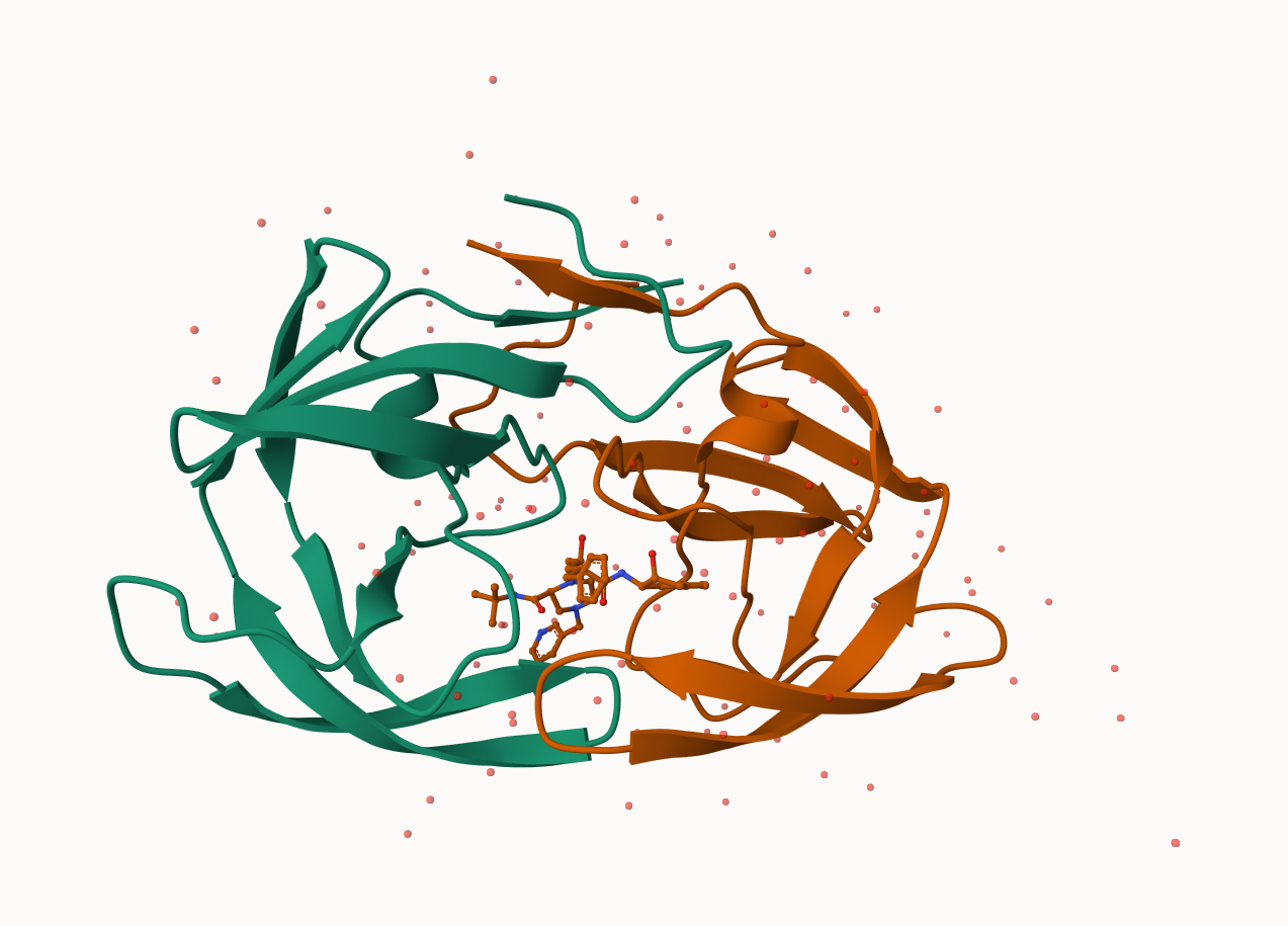
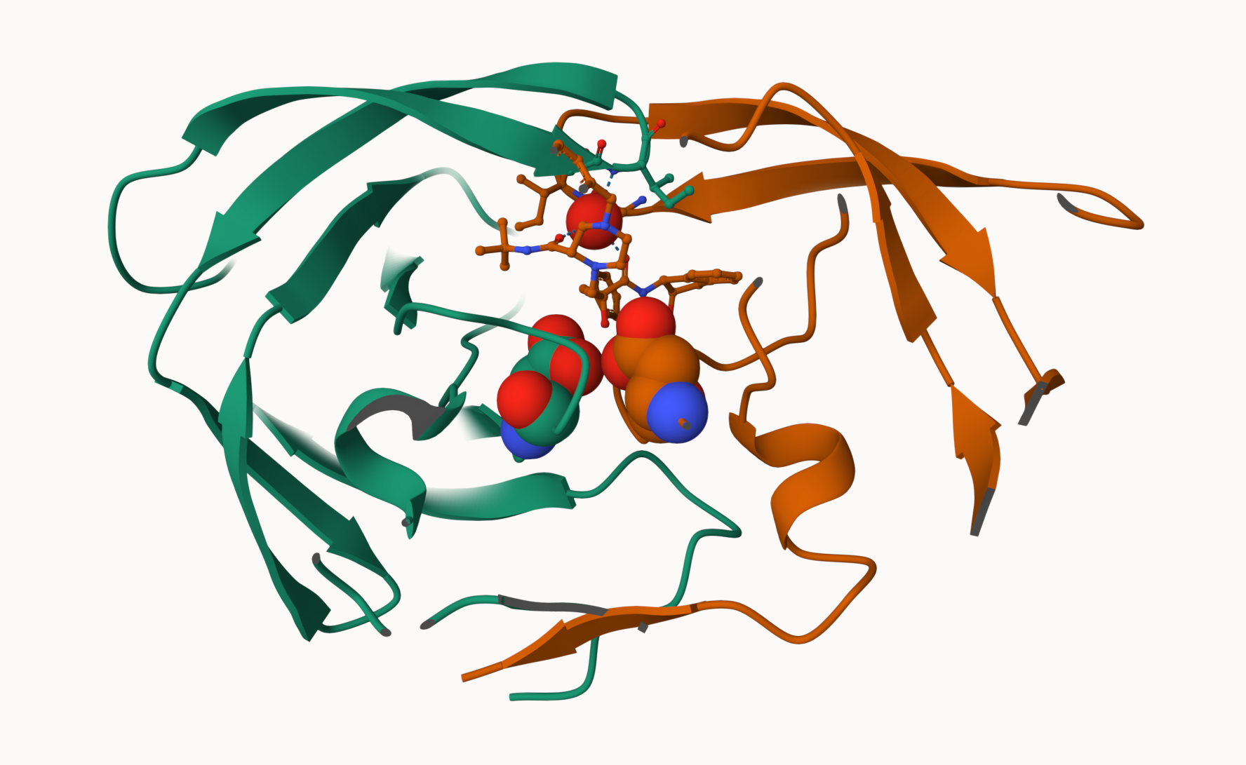
Let’s first use the Mol* viewer to explore this structure

My first view of HIV-Pr

PDB objects in R

head(hiv$atom)
  type eleno elety  alt resid chain resno insert      x      y     z o     b
1 ATOM     1     N <NA>   PRO     A     1   <NA> 29.361 39.686 5.862 1 38.10
2 ATOM     2    CA <NA>   PRO     A     1   <NA> 30.307 38.663 5.319 1 40.62
3 ATOM     3     C <NA>   PRO     A     1   <NA> 29.760 38.071 4.022 1 42.64
4 ATOM     4     O <NA>   PRO     A     1   <NA> 28.600 38.302 3.676 1 43.40
5 ATOM     5    CB <NA>   PRO     A     1   <NA> 30.508 37.541 6.342 1 37.87
6 ATOM     6    CG <NA>   PRO     A     1   <NA> 29.296 37.591 7.162 1 38.40
  segid elesy charge
1  <NA>     N   <NA>
2  <NA>     C   <NA>
3  <NA>     C   <NA>
4  <NA>     O   <NA>
5  <NA>     C   <NA>
6  <NA>     C   <NA>

Extract the sequence

pdbseq(hiv)
  1   2   3   4   5   6   7   8   9  10  11  12  13  14  15  16  17  18  19  20 
"P" "Q" "I" "T" "L" "W" "Q" "R" "P" "L" "V" "T" "I" "K" "I" "G" "G" "Q" "L" "K" 
 21  22  23  24  25  26  27  28  29  30  31  32  33  34  35  36  37  38  39  40 
"E" "A" "L" "L" "D" "T" "G" "A" "D" "D" "T" "V" "L" "E" "E" "M" "S" "L" "P" "G" 
 41  42  43  44  45  46  47  48  49  50  51  52  53  54  55  56  57  58  59  60 
"R" "W" "K" "P" "K" "M" "I" "G" "G" "I" "G" "G" "F" "I" "K" "V" "R" "Q" "Y" "D" 
 61  62  63  64  65  66  67  68  69  70  71  72  73  74  75  76  77  78  79  80 
"Q" "I" "L" "I" "E" "I" "C" "G" "H" "K" "A" "I" "G" "T" "V" "L" "V" "G" "P" "T" 
 81  82  83  84  85  86  87  88  89  90  91  92  93  94  95  96  97  98  99   1 
"P" "V" "N" "I" "I" "G" "R" "N" "L" "L" "T" "Q" "I" "G" "C" "T" "L" "N" "F" "P" 
  2   3   4   5   6   7   8   9  10  11  12  13  14  15  16  17  18  19  20  21 
"Q" "I" "T" "L" "W" "Q" "R" "P" "L" "V" "T" "I" "K" "I" "G" "G" "Q" "L" "K" "E" 
 22  23  24  25  26  27  28  29  30  31  32  33  34  35  36  37  38  39  40  41 
"A" "L" "L" "D" "T" "G" "A" "D" "D" "T" "V" "L" "E" "E" "M" "S" "L" "P" "G" "R" 
 42  43  44  45  46  47  48  49  50  51  52  53  54  55  56  57  58  59  60  61 
"W" "K" "P" "K" "M" "I" "G" "G" "I" "G" "G" "F" "I" "K" "V" "R" "Q" "Y" "D" "Q" 
 62  63  64  65  66  67  68  69  70  71  72  73  74  75  76  77  78  79  80  81 
"I" "L" "I" "E" "I" "C" "G" "H" "K" "A" "I" "G" "T" "V" "L" "V" "G" "P" "T" "P" 
 82  83  84  85  86  87  88  89  90  91  92  93  94  95  96  97  98  99 
"V" "N" "I" "I" "G" "R" "N" "L" "L" "T" "Q" "I" "G" "C" "T" "L" "N" "F" 
chainA_seq <- pdbseq(trim.pdb(hiv, chain="A"))

I can interactively view these PDB objects in R with the new bio3dview package. This is not yet on CRAN.

To install this I can setup pak package and use it to install bio3dview from GitHub. In my console I first run

install.packages(“pak”)

pak::pak("bioboot/bio3dview")
ℹ Loading metadata database
✔ Loading metadata database ... done
 
ℹ No downloads are needed
✔ 1 pkg + 40 deps: kept 34 [4.8s]
library(bio3dview)

view.pdb(hiv)

Change some settings

sel <- atom.select(hiv, resno=25)

view.pdb(hiv, highlight = sel,
         highlight.style = "spacefill",
         colorScheme = "chain",
         col=c("blue","orange"),
         backgroundColor = "pink")

Predict protein flexibility

We can run a bioinformatics calculation to predict protein dynamics - i.e. functional motions.

We will use the nma() function”:

adk <- read.pdb("6s36")
  Note: Accessing on-line PDB file
   PDB has ALT records, taking A only, rm.alt=TRUE
adk

 Call:  read.pdb(file = "6s36")

   Total Models#: 1
     Total Atoms#: 1898,  XYZs#: 5694  Chains#: 1  (values: A)

     Protein Atoms#: 1654  (residues/Calpha atoms#: 214)
     Nucleic acid Atoms#: 0  (residues/phosphate atoms#: 0)

     Non-protein/nucleic Atoms#: 244  (residues: 244)
     Non-protein/nucleic resid values: [ CL (3), HOH (238), MG (2), NA (1) ]

   Protein sequence:
      MRIILLGAPGAGKGTQAQFIMEKYGIPQISTGDMLRAAVKSGSELGKQAKDIMDAGKLVT
      DELVIALVKERIAQEDCRNGFLLDGFPRTIPQADAMKEAGINVDYVLEFDVPDELIVDKI
      VGRRVHAPSGRVYHVKFNPPKVEGKDDVTGEELTTRKDDQEETVRKRLVEYHQMTAPLIG
      YYSKEAEAGNTKYAKVDGTKPVAEVRADLEKILG

+ attr: atom, xyz, seqres, helix, sheet,
        calpha, remark, call
m <- nma(adk)
 Building Hessian...        Done in 0.012 seconds.
 Diagonalizing Hessian...   Done in 0.274 seconds.
plot(m)

Generate a “trajectory” of predicted motion

mktrj(m, file="ADK_nma.pdb")
view.nma(m)